Movies
Spider Development
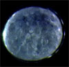
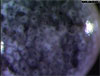
| 1 | Contraction | View Movie Contraction in Cheiracanthium mildei, side view. Elapsed time 6 hr. Movie courtesy of Bryce Cragg ’08. |
 |
|
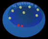
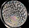
| 2 | Animation | View Animation Top of germ disc has been removed and cells are visible along cut edge. Top of yolk is dark blue. Cells ingress through the blastopore onto the yolk mass. Internalized cells become mesendoderm (yellow) or cumulus (red). Animation courtesy of Tony Moreno. |
 |
|
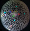
| 3 | Canonical | View Movie Normal development of Cheiracanthium mildei from nuclear division through hatching. Cumulus migrates to the left. Elapsed time 6.6 days. Movie courtesy of Christine Bates ’10 and Marion Burrill ’09. |
 |
|
| 4 | Cumulus in Zygiella | View Movie Cumulus formation and migration (gastrulation I) in Zygiella. Cumulus cells are colored red; some cells that will ingress during gastrulation II are in different colors. Cells were traced frame-by-frame, but movie shows one-hr intervals only. Posterior down. Elapsed time 20 hr. Movie courtesy of Emily Vance ’05. |
 |
|
| 5 | Primitive Plate in Zygiella | View Movie Primitive plate formation (gastrulation II) in Zygiella. Some ingressing cells are colored. Movie starts at cumulus formation and runs through gastrulation II. Cells were traced frame-by-frame, but movie shows one-hr intervals only. Posterior down. Elapsed time 24 hr. Movie courtesy of Emily Vance ’05. |
 |
|
| 6 | Gast I, II, III | View Movie Gastrulation I, II, and III (Stages C–H) in Zygiella. Note movement of internalized cells from center to periphery during gastrulation II. Cumulus migrates toward lower left. Elapsed time 45 hr. Movie courtesy of Emily Vance ’05. |
 |
|
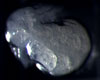
| 7 | Inversion in Zygiella | View Movie Inversion in Zygiella. Note that the rows of leg sockets move away from each other. Elapsed time 54 hr. Movie courtesy of Crystal Chaw ’02. |
 |
|
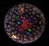
| Gastrulation in Latrodectus | View movie. Cumulus cells (red and pink) internalize first at the central blastopore. Yellow and orange cells internalize later to form generalized mesendoderm. Yellow cells at germ disc rim will internalize later. Blue cells are examples of cells known to persist in the superficial layer. Elapsed time 21 hr. Movie courtesy of Jennifer Turner '12. |
 | |
| ( Visit the spider development research page. ) | ||
If you have problems getting the movie to load, you may not have the QuickTime plug-in properly installed. Go to http://quicktime.apple.com to get assistance and information.